Overview: Learn about the different types of mammograms and when to consult your provider about breast symptoms.
A mammogram is the gold standard in breast imaging to look at breast tissue as this can detect breast cancer. But did you know there are different types of mammograms? Read the key differences between a screening and diagnostic mammogram:
Difference between screening and diagnostic mammograms
Screening mammograms
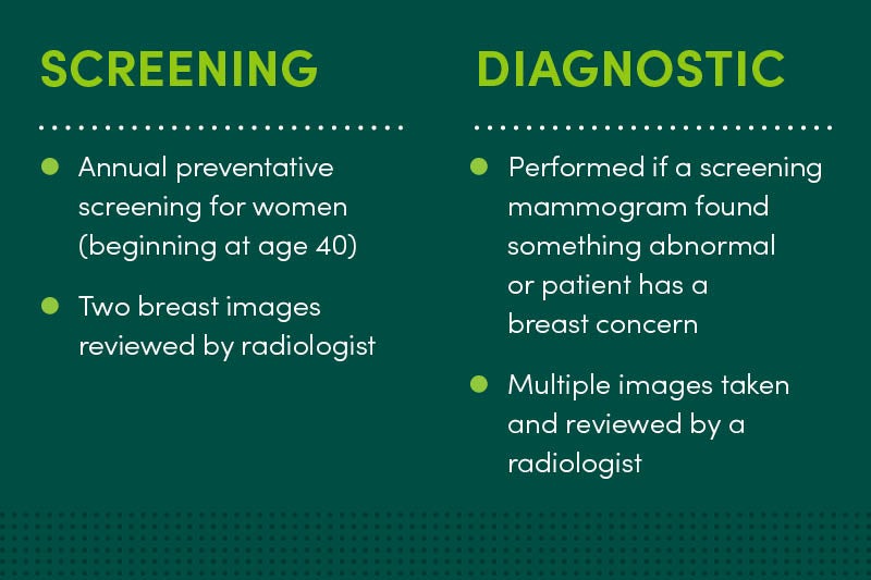
Screening mammograms are annual preventive screenings for women age 40 years or older with no symptoms of breast cancer. Two images of each breast are taken and reviewed by a radiologist after the exam.
Diagnostic mammograms
Diagnostic mammograms are performed if a screening mammogram found something abnormal or if someone is experiencing a breast concern or symptom. Multiple images are taken by a technologist and reviewed by a radiologist.
Watch for these symptoms:
- Lumps
- Pain in one or both breasts
- Nipple discharge
- Thickening of the skin on your breasts
- Swelling
- Redness
- Any changes in the size or shape of your breasts
Why would I need additional breast imaging?
During either a screening mammogram or diagnostic mammogram, your radiologist may determine that additional imaging is needed. This can include an ultrasound, MRI or image-guided breast biopsy.
Ultrasounds use sound waves to produce an echo and create images of breast tissue to show if an area in the breast is a cyst filled with fluid or a solid mass. Usually, a cyst isn’t cancerous, but a mass could lead to a cancerous tumor.
MRI creates a 3D image that provides a clear and detailed image of the breast. MRIs are recommended for women who have dense breasts. Having dense breasts can increase your breast cancer risk and make early detection more challenging.
A 3D mammogram (or tomosynthesis) is obtained when the machine moves in an arc around the breast to capture multiple images. A computer imaging program compiles the pictures into a highly detailed picture, with the ability to look at the inside of the breast, slice by slice. This gives the radiologist a more in-depth look at the breast.
Do mammograms hurt?
During a mammogram, your breasts are gradually pressed between two plates to keep them in place as the technician takes images. This may cause some temporary discomfort, but everyone is different.
When scheduling your mammogram appointment, try to schedule it around your period as most women experience sensitivity during that time. You can also take pain medication beforehand.
Learn more about what happens during a mammogram appointment.
If you have a breast concern, contact your primary care provider through MyChart to schedule a visit or contact your mammography office. Learn more about MercyOne personalized cancer care.
This blog was medically reviewed by a MercyOne provider.
